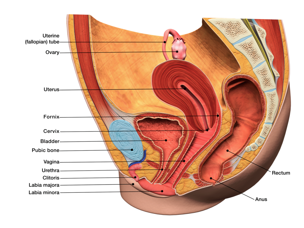
The vagina is an elastic muscular tube of 7 to 10 cm in length that extends from the vulva (female external genitalia) to the cervix of the uterus where it ends in an anterior and posterior fornix. The vaginal canal is positioned between the urethra and bladder anteriorly, and the rectum posteriorly.
The vaginal opening is in the posterior portion of the vulvar vestibule, behind the urethral opening. It is surrounded on either side by the labia minora medially and labia majora laterally. The vaginal canal has an outer fibrous adventitia, a middle layer of smooth muscle cells, and an inner layer of mucosa. The inner mucosal membrane has transverse folds called rugae.1

Vaginal lubrication is provided by the Bartholin’s glands near the vaginal opening and the cervix. The cells of the vagina are responsive to the hormone estrogen and contain receptors for this hormone, which produces a response that is crucial in the maintenance of the vaginal walls.1
Vaginal microbiota is a dynamic variable system, represented by a variety of bacteria, the vital activity and balance of which provides vaginal homeostasis. The importance of microbiota in the formation of vaginal health cannot be underestimated. The dominant constituent of the vaginal microbiota is lactobacillus.
Production of lactic acid, as a result of the vital activity of these bacteria, ensures the maintenance of the optimum low pH of the vaginal fluid, thus protecting from infections of the urogenital tract. The low pH of the vaginal fluid is also maintained by the active proton transport by the vaginal epithelium formed because of anaerobic glucose metabolism.2
2. Naumova, I. and Castelo-Branco, C. 2018 ‘Current treatment options for postmenopausal vaginal atrophy’, International Journal of Women’s Health. Volume 10, 387-395.

